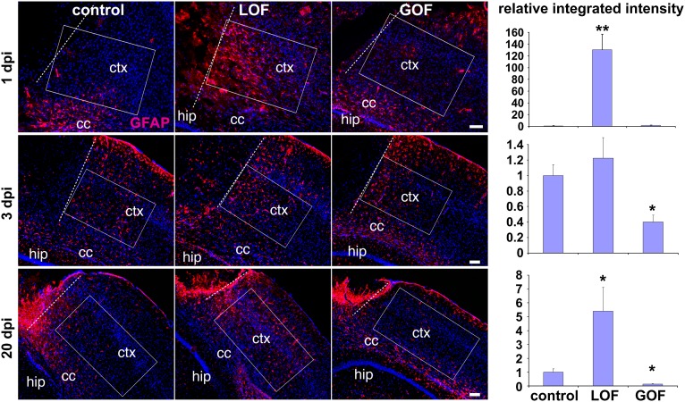
Fig. 6.
FGF signaling inhibits injury-induced astrocyte activation. Controls, LOF mutants, and GOF mutants were subjected to a cortical stab wound injury 3 wk after TM treatment. Representative images of coronal sections of GFAP immunostaining (red; Hoechst, blue) were taken at 1, 3, and 20 d post injury (dpi). Integrated intensity of GFAP staining was quantified in boxed areas parallel to the plane of the neocortex (ctx) immediately adjacent to the edge of the lesion (dotted line) for 1 and 3 d post injury, 130 μm from the lesion for 20 d post injury (to avoid the scar), and, in all cases, 100 μm from the corpus callosum (cc). Intensities were normalized to the measurement in the matching position of the contralateral hemisphere. The graphs represent the relative change of the integrated intensity to the mean measurement in controls, which was set as 1 (mean ± SEM; *P < 0.05 and **P < 0.001). hip, hippocampus. (Scale bars: 100 μm.)

