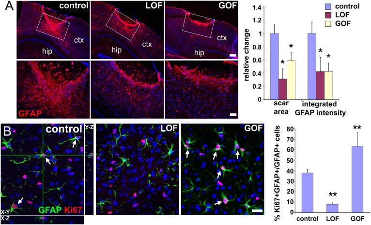
Fig. 7.
Aberrant FGF signaling reduces glial scar formation. (A) Immunostaining for GFAP in the neocortex (ctx) 20 d after a stab wound injury reveals significantly smaller scars not only in the GOF mutants, as expected, but also, surprisingly, in the LOF mutants. The area covered by scars and the integrated intensity of GFAP staining in the scar were measured in the boxed areas as described in Materials and Methods. For analysis, coronal sections were positionally matched between brains along the stab wound, which extends perpendicularly to the section along the anterior–posterior axis and which, because of the triangular shape of the blade, is deeper in more posterior areas. Changes are relative to the mean value obtained for controls (set as 1; mean ± SEM; *P < 0.05). hip, hippocampus. (Scale bars: Upper, 200 μm; Lower, 50 μm.) (B) Proliferation (Ki-67 staining) of reactive astrocytes (GFAP staining) around the lesion 3 d after injury is reduced in LOF mutants, but increased in GOF mutants, providing a potential explanation for why both mutants exhibit smaller scars. Colabeling for GFAP and Ki-67 (arrows) was quantified by confocal microscopy of 20-μm sections (mean ± SEM; **P < 0.001). (Scale bar: 25 μm.)

