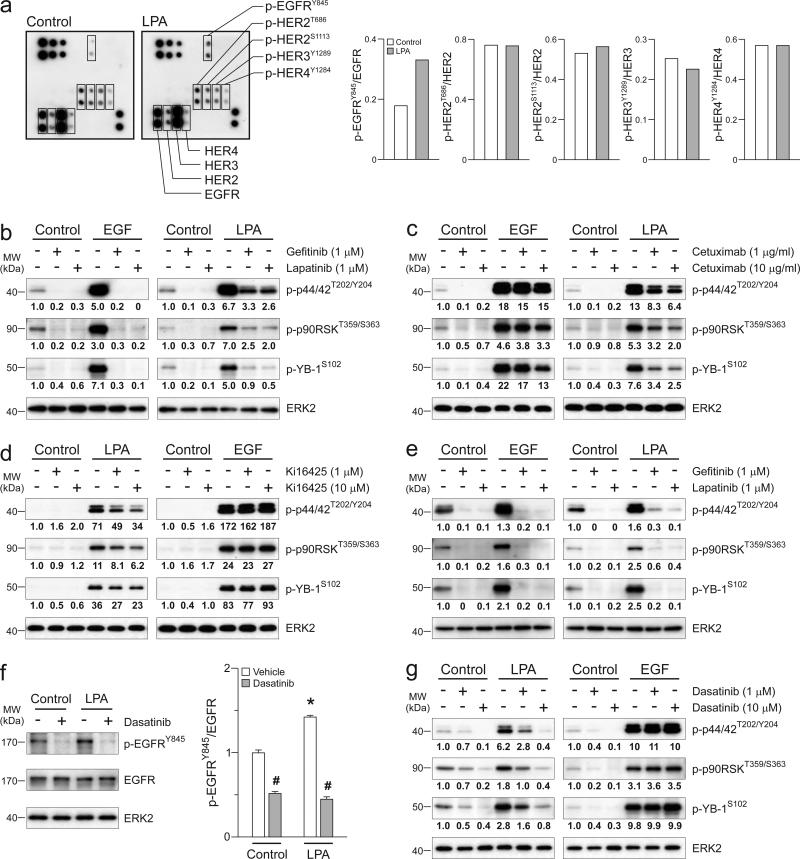
Figure 3.
LPA induces activation of p44/42MAPK/p90RSK/YB-1 pathway through SRC-mediated EGFR crosstalk. (a) CAOV3 cells were treated with LPA (10 μM) for 1h and phosphorylation levels of the EGFR/ERBB receptors were assessed using human EGFR family phosphorylation antibody array. Dot intensities were quantified on right. CAOV3 cells were treated with gefitinib or lapatinib (1 μM, b), Cetuximab (1 or 10 μM, c) or Ki16425 (1 or 10 μM, d) for 1h followed by LPA (10 μM) or EGF (10 ng/ml) for another 1h. (e) SKOV3 cells were treated with gefitinib or lapatinib followed by LPA or EGF similar to b. Western blot was performed in b-e. Band intensities of phospho-p44/42MAPK (p-44/42T202/Y204), phospho p90RSK (p-p90RSKT359/S363), phospho-YB-1 (p-YB-1S102) and YB-1 were quantified and normalized to the intensity of ERK2. (f) SKOV3 cells were treated with dasatinib (1 μM) for 1h followed by LPA (10 μM) for another 1h and Western blot was performed (left panel). Band intensities of phospho-EGFR (p-EGFRY845) were quantified and normalized to the intensity of EGFR (right panel). Data are means ± SEM, n = 3 independent preparations, *p < 0.05 compared to Control and #p < 0.05 compared to vehicle. (g) SKOV3 cells were treated with dasatinib and LPA similar to f and Western blot was performed with antibodies used in b-e. Western blot micrographs are representative of three independent preparations.

