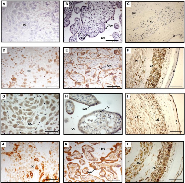
Figure 1.
Immunohistochemistry staining of term decidua basalis (A), chorionic villi (B) and fetal membranes (C) with AKR1B1 antibody (D–F), SLCO2A1 antibody (G–I), and PTGS2 antibody (J–L). Primary antibodies were omitted in (A–C) which served as controls. DC, decidual cells; Syn, syncytiotrophoblast; Cyt, cytotrophoblast; IVS, intervillous space; De, decidua parietalis; Ch, chorion; and A, amnion. Bar = 100 μm.

