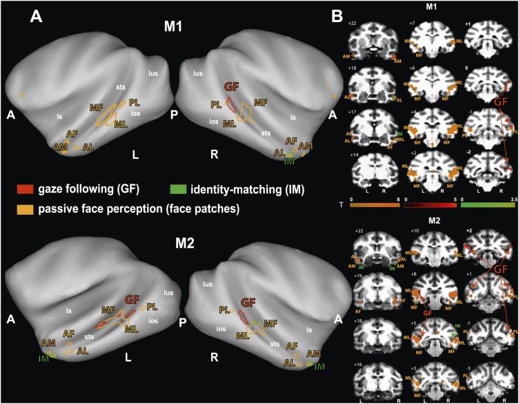
Figure 4. Comparison of the patterns of BOLD responses to 'gaze following' and 'identity matching' with the face patch BOLD pattern, delineated by the passive viewing of faces.
(A) Lateral views of the partially inflated hemispheres of monkeys M1 and M2 with borders of significant BOLD responses. Face patches (orange) based on 'faces vs nonfaces' contrast (p<0.05, uncorrected, 5 contiguous voxels) masked with an 'all non-scrambled vs all scrambled' objects' contrast (p<0.05 uncorrected). The red contours: significant BOLD contrasts for the 'gaze following vs identity matching' comparison (p<0.005, uncorrected, 5 contiguous voxels). The green contours: significant BOLD contrasts for the opposite, 'identity matching vs gaze following' comparison (p<0.05, uncorrected, 5 contiguous voxels). A = anterior, P = posterior, L = left, R = right, sts = superior temporal sulcus, ios = inferior occipital sulcus, lus = lunate sulcus, ls = lateral sulcus. (B) Coronal sections through the brains of monkeys M1 and M2 with corresponding significant BOLD contrasts from (A). The numbers in the left corners indicate the distance from the vertical interaural plane of each monkey (positive values anterior, neg. posterior) (L = left, R = right).


