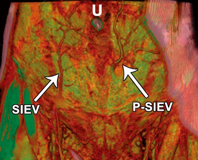
Figure 16.

Computed tomography angiogram (CTA) volume-rendered technique reconstruction of the anterior abdominal wall vasculature (anterior view), demonstrating a normal superficial inferior epigastric vein (SIEV) on the right and an anomalous ‘perforating’ SIEV (P-SIEV) on the left. U=Umbilicus (Reproduced with permission from: Rozen WM, Grinsell D, Ashton MW. The perforating superficial inferior epigastric vein: a new anatomical variant detected with computed tomographic angiography. Plast Reconstr Surg 2010;125:119e-20e).
