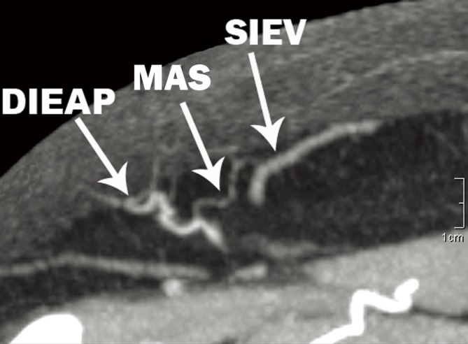
Figure 18.

Axial slice of an arterial phase computed tomographic angiogram (CTA), demonstrating the presence of a 1 mm macrovascular arteriovenous shunt (MAS) between a large deep inferior epigastric artery perforator (DIEAP) and the superficial inferior epigastric vein (SIEV) (Reproduced with permission from: Rozen WM, Chubb D, Ashton MW, et al. Macrovascular arteriovenous shunts (MAS): a newly identified structure in the abdominal wall with implications for thermoregulation and free tissue transfer. J Plast Reconstr Aesthet Surg 2010;63:1294-9).
