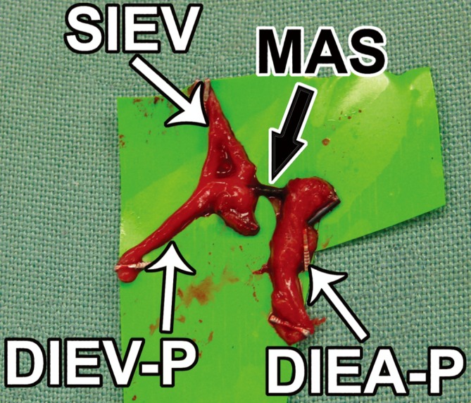
Figure 21.

The macrovascular arteriovenous shunt (MAS) is shown ex-vivo, after excision of the structure. Again, the MAS communication between a DIEA perforator (DIEA-P) and the SIEV is demonstrated, with an adjacent deep inferior epigastric vein perforator (DIEV-P) (Reproduced with permission from: Rozen WM, Chubb D, Ashton MW, et al. Images in plastic surgery: the anatomy of macrovascular arteriovenous shunts and implications for abdominal wall free flaps. Ann Plast Surg 2011;67:99-100).
