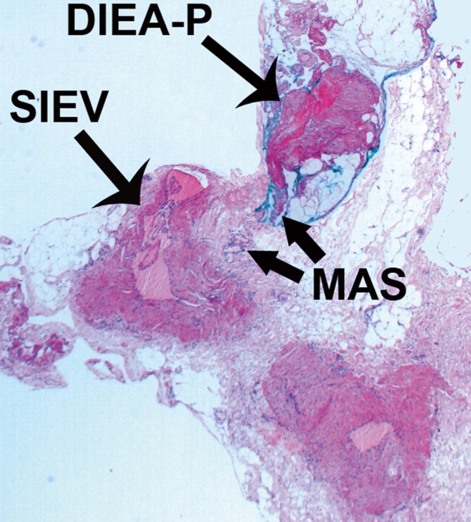
Figure 22.

Histological analysis with Hematoxylin and Eosin (H and E) stain of a transverse section through the macrovascular arteriovenous shunt (MAS) shown in Figures 1A and 1B. ‘Arterial’ features are seen on the arterial side of the shunt, with a thicker wall, particularly the media and thinner lumen, while ‘venous’ features on the venous side, with thinner wall and wider lumen. Scale as shown (30 times magnification) (Reproduced with permission from: Rozen WM, Chubb D, Ashton MW, et al. Images in plastic surgery: the anatomy of macrovascular arteriovenous shunts and implications for abdominal wall free flaps. Ann Plast Surg 2011;67:99-100).
