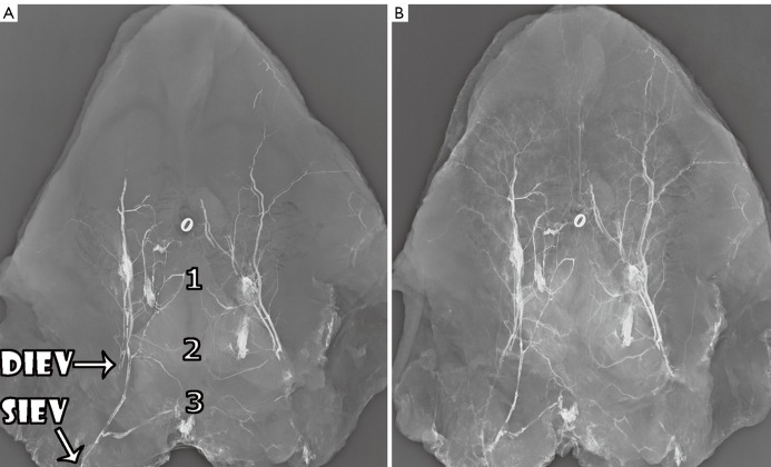
Figure 5.
A. Plain film venogram of the anterior abdominal wall, with deep inferior epigastric vein (DIEV) and superficial inferior epigastric vein (SIEV) and periumbilical communicating veins all injected with contrast. Three points of midline crossover in this specimen are highlighted; B. The deep inferior epigastric artery (DIEA) is also injected in the same specimen, confirming that the left image is a venogram, and demonstrating the concomitant nature of the veins. O = umbilicus (Reproduced with permission from: Rozen WM, Pan WR, Le Roux CM, et al. The venous anatomy of the anterior abdominal wall: an anatomical and clinical study. Plast Reconstr Surg 2009;124:848-53).

