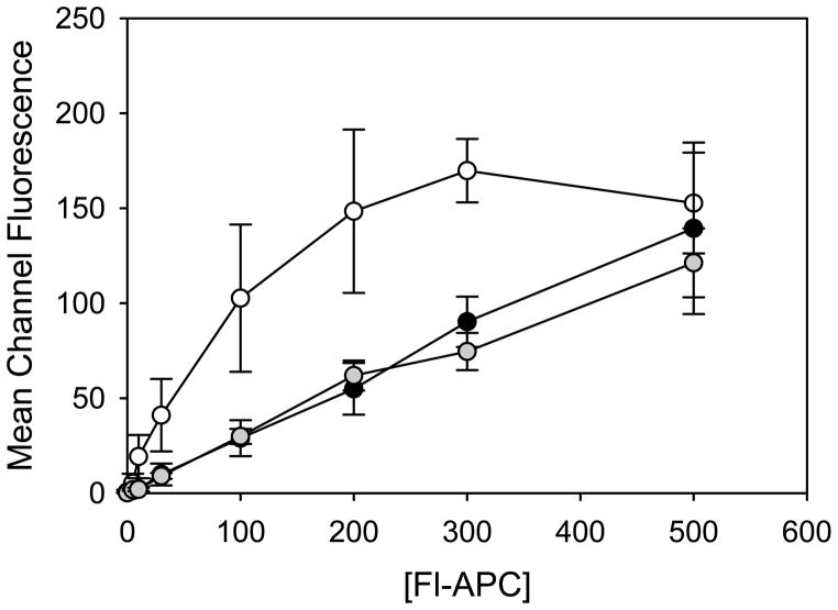
Figure 5. Binding of Fl-APC to WT EPCR or EPCR variants expressed on HEK293 cells.
The binding of fluorescently-labelled APC (Fl-APC) to EPCR variants was assessed by flow cytometry as described under “Materials and Methods”. The values correspond to the mean and the S.E. of 3 separate experiments. Symbols: WT EPCR (white circles); EPCR Val170Leu (gray circles); EPCR Arg96Cys (black circles).

