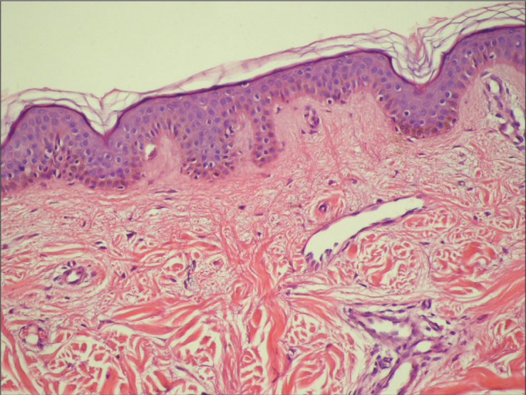
Figure 2.

Biopsy specimens from the upper back of the Case 1 demonstrate the edematous changes seen with scleredema. In the dermis, there are abundant thickened collagen bundles separated by clear spaces. The collagen fibers are also irregularly arranged (H&E, original magnification ×100)
