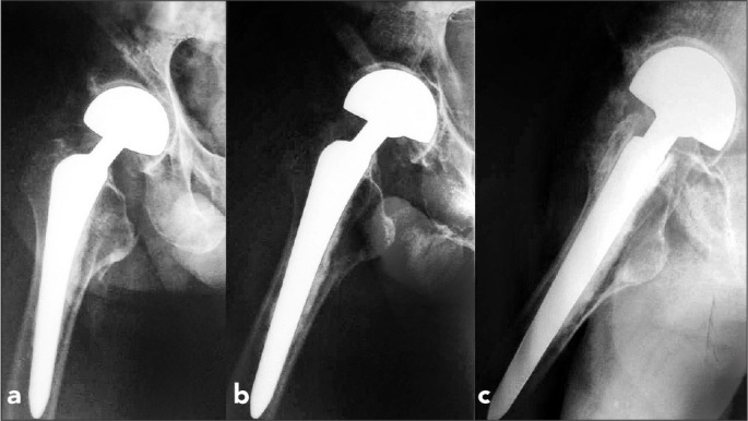
Figure 2.

a–c. Twenty-four-month postoperative radiographs of a 76-year-old man. Radiograph taken in adduction position (a). Radiograph taken in abduction position (b). Radiograph taken in lateral position (c). All movement ensued at the inner bearing (metal/polyethylene) surface (Type A). Hip motion did not lead to alteration of the outer articular surface position
