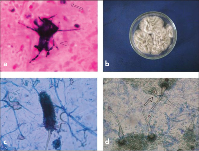
Figure 2.

a–d. Leucocytes (arrow) and hyphae (arrowhead) in direct microscopic analysis of sputum (Gram staining) (a), colony appearance in Saboraud Dextrose Agar (SDA; 42°C, 3 days) (b), conidiospore structure of the strain in slide culture (SDA, 25°C) (c), and microscopic appearance of the branched conidiophores (arrow) in SDA (25°C) (d)
