
Fig. 2.
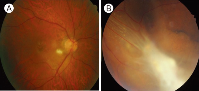
Fundus photographs of retinal granuloma in a 67- (A) and 31-year-old male (B) patients with ocular toxocariasis. (A) Posterior pole granuloma appears as an oval, white lesion in the posterior pole of the retina. (B) Peripheral granuloma presents with an amorphous whitish mass with tractional membrane and retinal detachment.