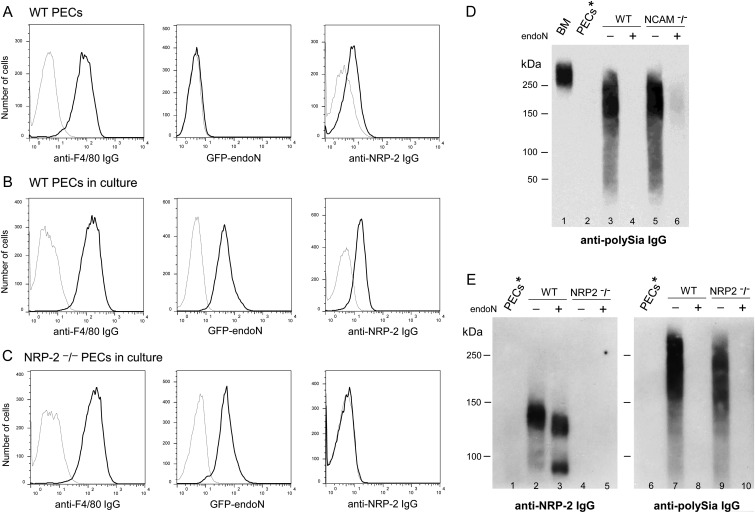
Fig. 7.
Peritoneal macrophages lose expression of polySia on the cell surface but can re-express polySia as a modification of NRP-2 and another protein(s). Peritoneal macrophages were isolated 72 h after injection of thioglycollate into the peritoneal cavity of wild-type, NCAM−/− and NRP-2−/− mice. Freshly-isolated PECs from wild-type mice were evaluated by flow cytometry after staining with anti-F4/80 and -NRP-2 IgGs and with GFP-endoN: thin lines—isotype IgGs or cells that were treated with endoN prior to staining with GFP-endoN, bold line—staining of cells with specific IgGs or GFP-endoN (A). After isolation from the peritoneal cavity, PECs were grown in culture in RPMI/10% FCS for 24 h, were harvested and were stained with F4/80 and NRP-2 IgGs and with GFP-endoN (B: cultured PECs from wild-type mice; C: cultured PECs from NRP-2−/− mice). Immunoblot of proteins from lysates of unfractionated BM cells (5 μg, lane 1), freshly-isolated peritoneal macrophages (25 μg, lane 2), macrophages grown in culture for an additional 24 h from wild-type (5 μg, lanes 3 and 4) or NCAM−/− (5 μg, lanes 5 and 6) mice without (lane 3 and 5) or with (lane 4 and 6) digestion with endoN probed for polySia expression with mAb735 (D). Immunoblots of proteins from lysates of freshly-isolated peritoneal macrophages (30 μg, lanes 1 and 6), macrophages grown in culture for an additional 24 h from wild-type (30 μg, lanes 2 and 3; 5 μg, lanes 7 and 8) or NRP-2−/− (30 μg, lanes 4 and 5; 5 μg, lanes 9 and 10) mice without (lanes 2, 4, 7 and 9) or with (lanes 3, 5, 8 and 10) digestion with endoN probed with antibody to NRP-2 (lanes 1–5) or to polySia (lanes 6–10) (E).

