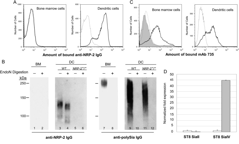
Fig. 8.
Murine bone marrow-derived DCs express polysialylated NRP-2. Freshly isolated murine BM cells and BM-derived DCs were stained with anti-NRP-2 IgG and analyzed by flow cytometry (A): thin, dotted line—isotype IgG; bold line—anti-NRP-2 IgG. Lysates of BM cells from wild-type mice (lanes 1, 2, 7 and 8) or DCs from wild-type (lanes 3, 4, 9 and 10) or NRP-2−/− (lanes 5, 6, 11 and 12) mice that were mock treated (lanes 1, 3, 5, 7, 9, 11) or endoN treated (lanes 2, 4, 6, 8, 10 and 12) were separated by 8% SDS–PAGE and analyzed by immunoblot using anti-NRP-2 IgG (left panel) or mAb 735 (right panel) (B). Bone marrow cells and BM-derived DCs were stained with anti-polySia mAb 735 and analyzed by flow cytometry (C): thin, dotted line—isotype IgG, shaded region—endoN-treated BM cells stained with mAb 735; bold line—mock-endoN-treated cells stained with mAb 735. The relative amount of RNA encoding ST8 SiaII and ST8 SiaIV in BM cells (open bars) and DCs (shaded bars) was determined by semi-quantitative PCR (D). The amount of RNA encoding each enzyme in BM cells was normalized to one and the fold change was in relation to the change in expression of 18S rRNA.

