Figure 7.
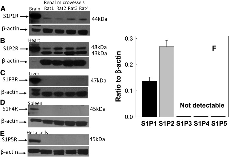
Protein expression of S1P receptors in isolated rat preglomerular microvessels. Representative Western blot images for S1P1 (A), S1P2 (B), S1P3 (C), S1P4 (D), and S1P5 (E) receptor expression in isolated preglomerular microvessels. Homogenates from brain, heart, liver, spleen, and HeLa cells are used as positive control tissues for S1P1, S1P2, S1P3, S1P4, and S1P5 receptor expression, respectively. β-Actin serves as a loading control and is shown in the bottom of each panel. Densitometry analysis of S1P receptor protein expression in isolated preglomerular microvessels is summarized in F (n=4 rats for each receptor studied). Data are expressed as the mean±SEM.

