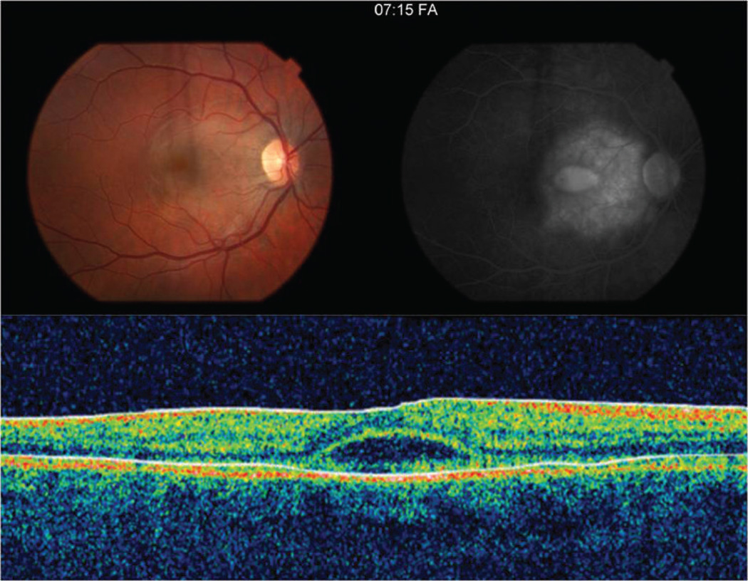
Figure 1.
The color fundus photograph of the affected right eye (A) one day after symptom onset demonstrates an irregular, circular area of grey-white discoloration in the macula. (B) Late-phase fluorescein angiogram image shows a large area of intense subretinal hyperfluorescence consistent with leakage and a smaller central area of pooling coinciding with an exudative detachment of the neurosensory retina. (C) TD-OCT shows a small subfoveal neurosensory detachment with hyperreflective debris at the apical side of the retinal pigment epithelium.

