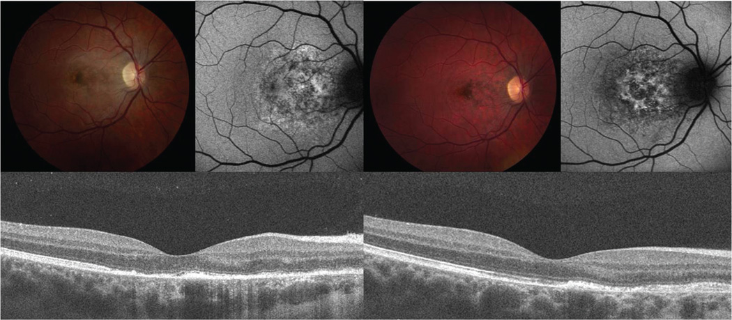
Figure 3.
Color fundus photograph, FAF, and SD-OCT findings in patient 1 at one week (A) and two months (B) after symptom onset. There is a loss of the grey-white discoloration and an increase in retinal pigment hyperplasia later in the disease course. The FAF shows less stippled autofluorescent areas (A, upper right) that evolve to a more stellate shaped autofluorescence with loss of background autofluorescence (B, upper right). SD-OCT shows hyporeflectivity of the outer photoreceptor layer (A, arrows), that are partially restored (B, arrowheads) at the twomonth follow-up visit. Note that the external limiting membrane remains intact in both A and B, yet the apical debris on the RPE has diminished.

