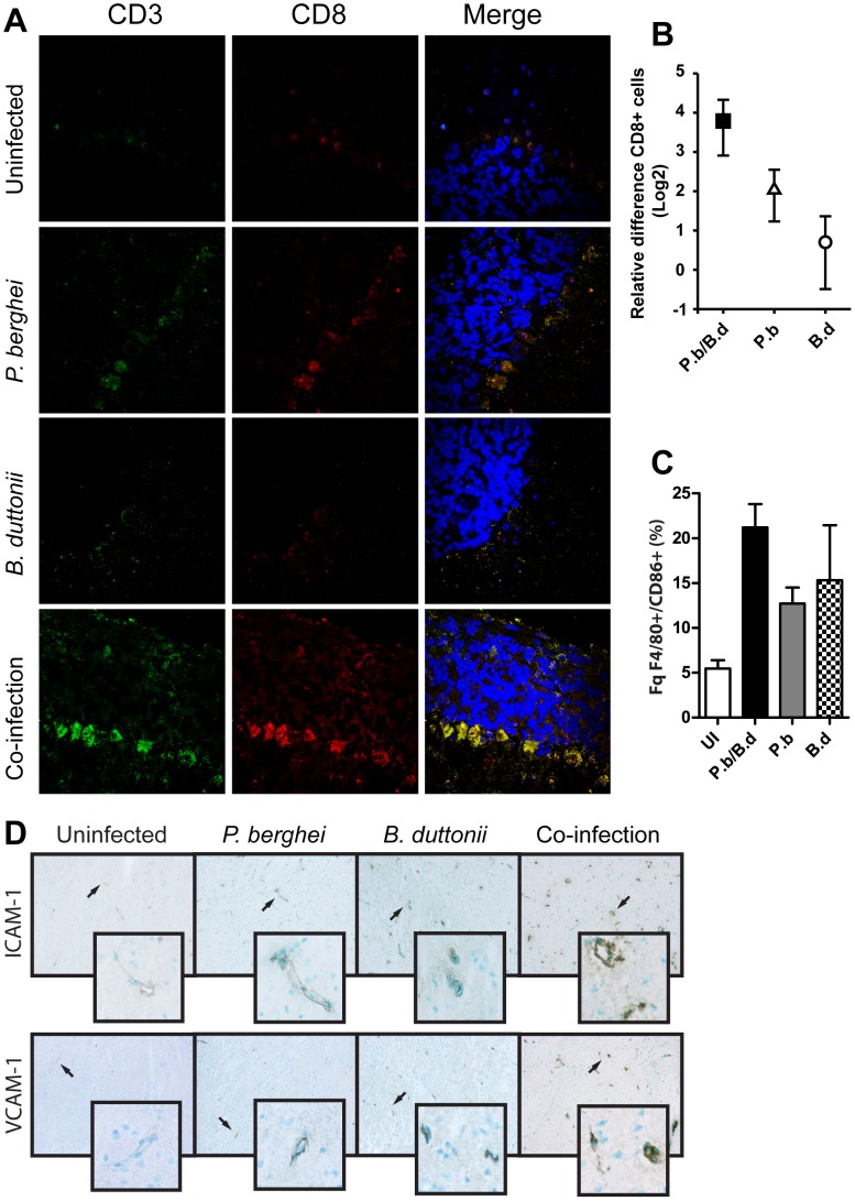
Figure 2. The brain in co-infected mice contain high amounts of CD8+ cells and activated MΦs.
Brains from mice infected with P. berghei, B. duttonii or both as per M&M were collected day 5 p.i. (N = 6 per group), experiments performed in duplicate (A) Cryo-sectioned tissue double stained for CD3 and CD8 positive cells and DNA visualized with DAPI. Representative regions in the frontal cortex were imaged by confocal laser microscopy. Tissue stained in absence of primary CD3 and CD8 antibodies served as negative controls. (B) The relative fold-change differences in absolute numbers of CD8+ cells. (C) The percentage frequency of CD86+ MΦs in the total pool of F4/80+ cells extracted from the brains (N = 6 per group, experiment performed in duplicate). (D) Cryo-sectioned brain tissue, immuno-stained for ICAM and VCAM.

