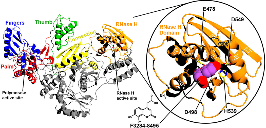
Figure 1. Ribbon diagram of the structure of full-length RT.
RT is a heterodimer that consists of p66 (color) and p51 (gray) subunits. The two enzymatic active sites and the subdomains of the p66 subunit are labelled. Right inset: A closer view of the isolated RNase H domain of the current structure, with F3284-8495 bound at the active site. Selected alpha-helices and beta-sheets are labelled based on ref. 52. The chemical structure of F3284-8495 is shown at the bottom center of the figure.

