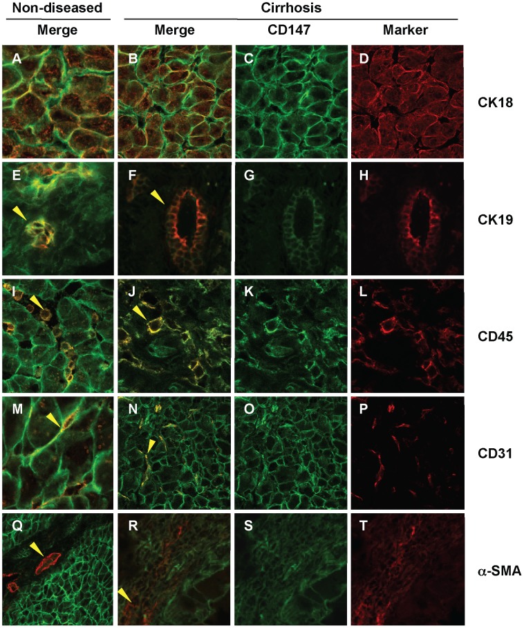
Figure 6. Co-localisation of CD147 and Liver Cell Markers in Human Liver Tissue.
Liver sections were stained with liver cell makers (red) including CK18 (Panels A, B and D), CK19 (Panels E, F and H), CD45 (Panels I, J and L), CD31 (Panels M, N and P) and α-SMA (Panels Q, R and T) as well as CD147 (green, all except D, H, L, P and T). CK-19 positive bile ducts (Panels E, F and H) co-localise with CD147 (Arrowheads Panel E and F) in both non-diseased and cirrhotic tissue. Similarly, CD45 positive leukocytes (Panels I, J and L) co-localise with CD147 in both non-diseased and cirrhotic tissue (Arrowheads in Panel I and J). Further, CD31 endothelial cells (Panels M, N and P) co-localise with CD147 (Panel M and N) in both non-diseased and cirrhotic tissue. Finally, in the α-SMA positive series of images (Panels Q, R and T) vascular structures are seen stained as indicated by the arrowhead in Panel Q and HSC in fibrotic septa in cirrhosis (Arrowhead in Panel R). Importantly, no co-localisation of α-SMA and CD147 was seen in cirrhosis (Panel R). Merged images show co-localisation of CD147 with the liver cell markers (yellow). Magnification 63×.

