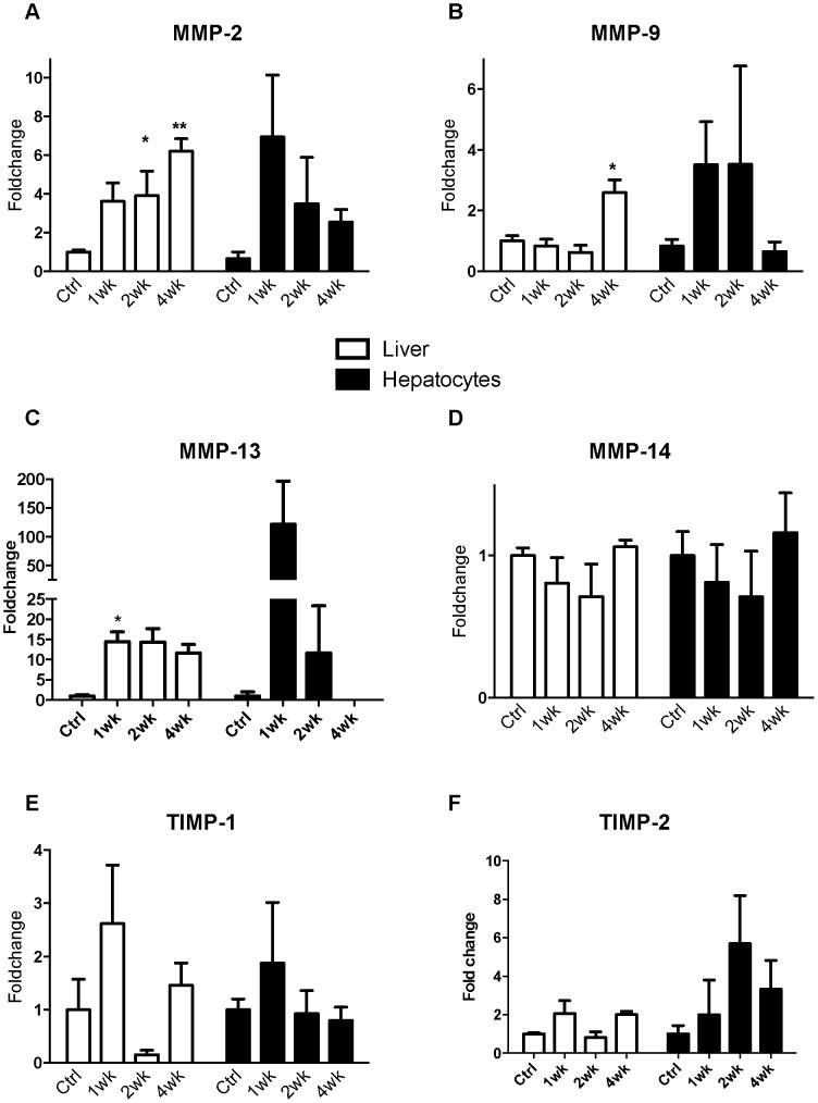
Figure 8. MMP and TIMP expression in whole liver and primary hepatocytes from CCl4 induced liver injury.
Quantitative PCR of MMP and TIMP expression in whole liver and isolated primary hepatocytes. Cirrhosis was induced in a mouse model of liver injury (C57bl/6) with CCl4. RNA extracted from primary hepatocytes as well as whole liver were assessed at commencement of injury and weeks 1, 2 and 4. The expression of MMP-2 (Panel A), MMP-9 (Panel B), MMP-13 (Panel C), MMP-14 (Panel D), TIMP-1 (Panel E) and TIMP-2 (Panel F) was assessed by quantitative PCR (n = 4 per group, data expressed as mean and SEM. *p<0.05 and **p<0.01 relative to untreated).

