
Fig. 1.
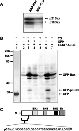
NRP-154 cells display cleavage of Bax during apoptosis. A: Western blot with anti-Bax (Δ21 antibody) reveals proteolysis of Bax in NRP-154 cells undergoing transforming growth factor (TGF)-β-induced apoptosis. Cells were treated with 10 ng/ml TGF-β for 48 h. B: Hela cells were transfected with either green fluorescent protein (GFP, lane 1) or GFP-Bax (lanes 2–7). Three hours after transfection, media were changed to either growth media without thapsigargin (TG, lanes 2–4) or with 2 μM TG (lanes 5–7). Transfected cells were treated with additional caspase inhibitor Oph-109 (lanes 3, 4, 6, and 7) to allow for cell survival until the end of the assay. Calpain inhibitors (ALLN and E-64d) were applied to lanes 4 and 7. Total cell lysates were collected 18 h after treatment and probed with anti-GFP (lane 1) or anti-Bax 6A7 (lanes 2–7). C: schematic diagram illustrates that a 32 amino-acid peptide (p3Bax) is generated from Bax (p21) via cleavage by calpain.