
Fig. 1.
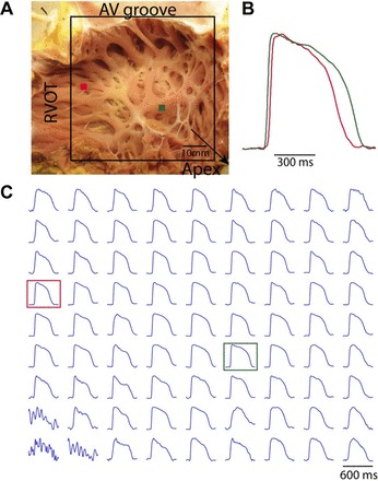
Representative right ventricular (RV) free wall preparation and optical action potentials (AP). A: RV free wall preparation and mapping field of view (FOV). B: close-up view of two AP recordings. C: representative optical AP from an evenly spaced array of locations spanning the whole FOV. Red and green dots in A and squares in C correspond to recordings in B. RVOT, right ventricular outflow tract.