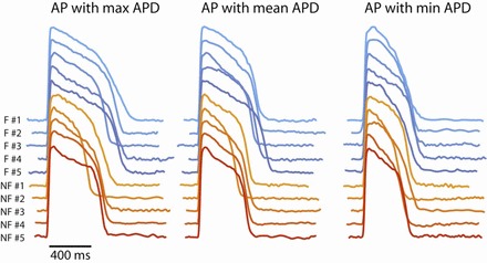
Fig. 3.

Representative AP recordings from individual failing (blue traces) and nonfailing (red traces) human hearts at the pacing cycle length (CL) of 2,000 ms. Left, AP recordings with the maximum APD in the FOV. Middle, the AP recordings with the mean APD in the FOV. Right, the AP recordings with the minimum APD in the FOV.
