
Fig. 6.
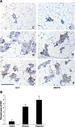
A: immunohistochemical staining (brown color as indicated by arrows) of the macrophage marker ED1 (left) and the matrix metallopeptidase MMP8 (right) in the mammary gland of rats at day 49 after a full-term pregnancy (P) or exogenous hormone treatment (H) compared with the control (C) rats. One representative staining is shown for each group. Scale bar = 200 μm. B: graphs representing the percentages of ED1-stained areas per unit of parenchyma and stroma. Different lowercase letters represent P < 0.01.