
Fig. 6.
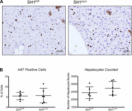
Hepatocyte proliferation after long-term PPARα treatment. Sirt1F/F and Sirt1ΔLiv mice were treated with WY for 3 mo. Immunohistochemistry was performed on formalin-fixed paraffin-embedded liver tissue sections for ki67, a marker of proliferation. A: representative images (×200) for ki67 staining in Sirt1F/F and Sirt1ΔLiv mice. B: %Ki67-positive cells counted for each mouse. No statistically significant difference in %ki67-positive cells or total nuclei was observed.