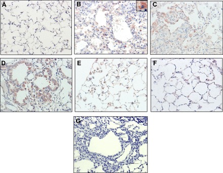
Fig. 6.

Immunolocalization of NFκB2 in lung tissue during the time course of bleomycin-induced lung fibrosis. Representative photomicrographs of immunohistochemical staining performed with specific antibody against NFκB2 at 1 (B), 4 (C), 8 (D), 12 (E), and 16 (F) wk after bleomycin instillation. Saline-instilled mouse lung is shown in A, and negative control of 8 wk postbleomycin in which the primary antibody was omitted is shown in G. Immunoreactive protein was observed in alveolar epithelial cells (C and D) and macrophages (B inset, E, and F). All sections were counterstained with hematoxylin. Scale bars indicate 50 μM.
