
Fig. 2.
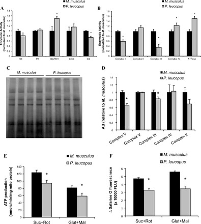
Activity of glycolytic and oxidative enzymes and electron transport chain complexes. A: skeletal muscle homogenates were used to test glycolytic enzyme activities: hexokinase (HK), pyruvate kinase (PK), and glyceraldehyde 3-phosphate dehydrogenase (GAPDH), and oxidative enzymes, cytochrome-c oxidase (COX), and citrate synthase (CS) (n = 6–12). B: activities of respiratory complex chains, including NADH dehydrogenase (complex I), succinate dehydrogenase (complex II), ubiquinone:cytochrome-c oxidoreductase (complex III), cytochrome-c oxidase (complex IV), and ATPase. Mitochondrial proteins were solubilized with dodecyl maltoside (1%), and membrane-enriched soluble proteins were then used to measure electron transport chain (ETC) activity either using a spectrophotometric assay or an in-gel assay, see details in materials and methods (n = 6–10). C: representative blue-native gel image of electron transport chain complexes. D: quantification of protein contents of ETC (n = 8). E: production of ATP was measured in isolated skeletal muscle mitochondria using a luciferase/luciferin assay kit as described in materials and methods (n = 6). F: mitochondrial membrane potential (ΔΨm) was measured using fluorescent safarine O dye. The quenching of safarine O fluorescence is proportional to the electrical potential across the mitochondrial inner membrane (n = 6). Black bar represents M. musculus, and gray bar represents P. leucopus. Data are expressed as means ± SE. *P < 0.05 compared with M. musculus.