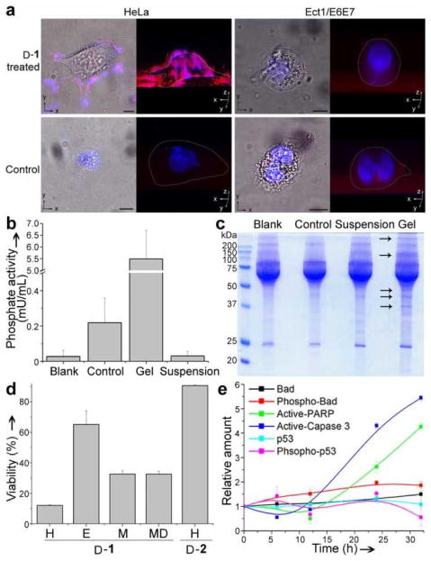
Figure 3.
The pericellular hydrogel/nanonets inhibit cancer cells. a) Overlaid images and 3D stacked z-scan images of Congo red and DAPI stained HeLa and Ect1/E6E7 cell treated by D-1 or just culture medium as control for 12 h. HeLa cells treated by D-1 at 280 μM; Ect1/E6E7 cells treated by D-1 at 560 μM. Scale bar = 10 μm. White dots outline the cells. b) Comparison of phosphate activity in blank medium (Blank), medium incubated with HeLa cells (Control), pericellular hydrogels on HeLa cells treated by D-1 at 560 μM (Gel), and the suspension medium of HeLa cells treated by D-1 at 560 μM (Suspension). c) SDS-PAGE showing the protein composition in the Blank, Control, Gel, and Suspension. Arrows point at protein bands that appear only in the lane of the Gel. d) Cell viabilities of HeLa (H), Ect1/E6E7 (E), MES-SA (M) and MES-SA/Dx5 (MD) cells treated by 280 μM of D-1 or HeLa cells treated by 280 μM of D-2 for 48 h. e) Change of relative amount of apoptosis signal molecules over time in HeLa cells treated by D-1 at 280 μM.

