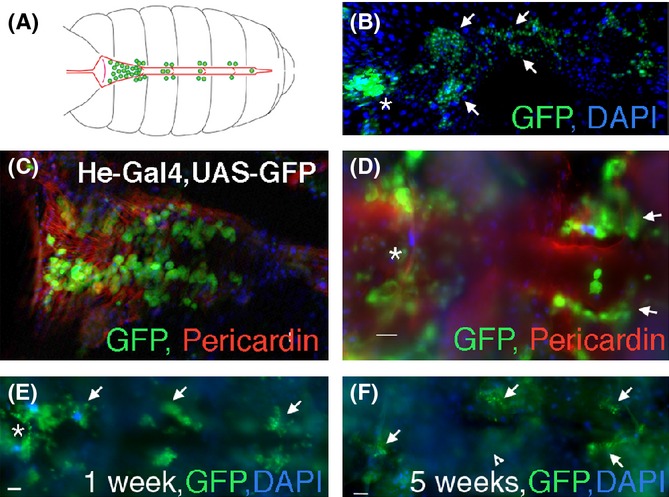
Figure 1.

Heart-associated hemocytes decline with age but not infection. (A) A schematic of the abdominal dorsal vessel (red) and associated blood cells (hemocytes, green). Hemocytes are enlarged for clarity. Anterior is to the left in this and subsequent panels. Many circulating hemocytes cluster in the first aortic chamber of the dorsal vessel (left), while some can be seen in the other chambers. Adherent hemocytes localize just outside the dorsal vessel and along the body wall. (B) Projection of multiple optical sections of a dissected dorsal vessel and associated blood cells from a 1-week-old female, marked by GFP (green) driven by Hemese-Gal4 (He-Gal4). Many circulating hemocytes are seen inside the first chamber (asterisk), and clusters of sessile hemocytes are found associated laterally along the dorsal vessel. Arrows indicate clusters of hemocytes along the second and third chambers. DAPI labels all nuclei in blue. (C) An optical section of a dissected dorsal vessel from a 1-week-old female shows GFP-positive hemocytes (green, arrow) inside of the first chamber of the dorsal vessel, which is marked by staining with an antibody directed against Pericardin (red). (D) In a different optical section, heart-associated hemocytes are observed adhering outside of the first chamber (asterisk), and laterally next to the second chamber of the dorsal vessel (arrows). Two blood cells are seen in the heart. (E) A dorsal vessel from a 1-week-old He-Gal4, UAS-GFP female injected with Escherichia coli displays hemocytes inside the first chamber (asterisk), and clusters of hemocytes laterally along the dorsal vessel (arrows). (F) A dorsal vessel from a 5-week-old He-Gal4, UAS-GFP female injected with E. coli displays similar localization of hemocytes inside the first chamber (asterisk), and laterally along the dorsal vessel (arrows), although there are fewer blood cells. Arrowhead indicates an out-of-focus cluster of cells. All scale bars = 20 μm.
