
Figure 4. Engaged centrioles block luminal recruitment of SAS-6.
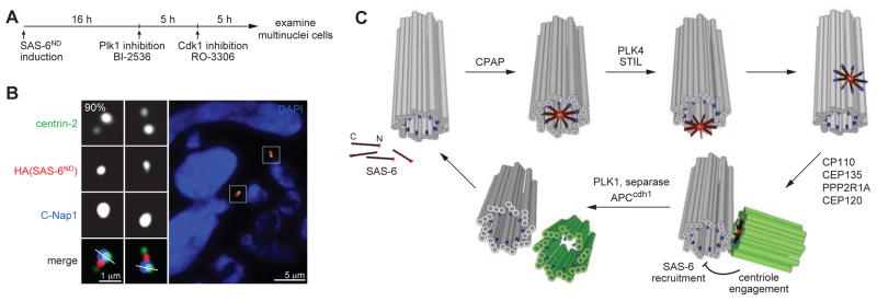
(A) Schematic outlining the generation of G1 cells with engaged centrioles as described previously (Tsou et al., 2009; Wang et al., 2011). SAS-6ND expression was induced in proliferating cells for 16 hours before adding BI-2536 to inhibit Plk1. Mitotic arrested cells were forced to exit mitosis 3 hours after Plk1 inhibition by Cdk1 inhibitor RO-3306 for 5 hours. Multinuclei cells, which were cells that had gone through mitosis in the absence of Plk1 activity, were examined for the centriolar localization of SAS-6 with indicated antibodies. n>26, N=3.
(B) SAS-6 recruitment to mother centriole lumen is inhibited by engaged centrioles. Immunofluorescence image of a G1 cell containing engaged centrioles with indicated antibodies.
(C) Model for a template-based mechanism of centriole duplication. SAS-6 dimers are individually recruited to the proximal lumen of mother centrioles, where they are organized by the surrounding geometry to undergo 9-fold symmetrical assembly. The assembled SAS-6 oligomer is then released from the lumen and forms the cartwheel that drives daughter centriole formation. Centriole engagement blocks re-recruitment of SAS-6 to the mother centriole lumen and thus prevents centriole re-duplication. Centriole disengagement and cartwheel removal at the end of mitosis allow the centrioles to be used as a template for their own biogenesis. Mother centriole (grey), daughter centriole (green), CPAP (blue), SAS-6 (red).