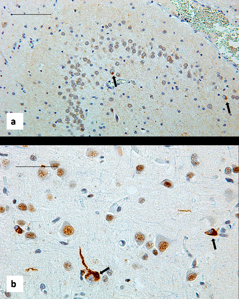
Figure 5.
Brain histology. Sections stained with antibody to total TDP-43 show scattered neurons with displacement of this protein from the nucleus to the cytoplasm, forming rounded or tangle like inclusion marked by black arrows. (a): patient IV-10, Dentate fascia of the hippocampus, bar 100 um. (b): patient IV-9 Temporal neocortex, bar 50 um.

