
Figure 3. Inhibition of PI3K signaling by BKM120 leads to the persistence of DNA damage.
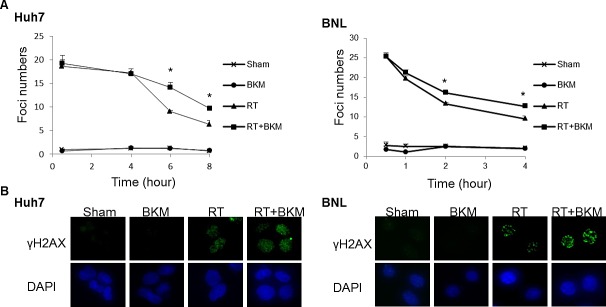
(A) The number of γ-H2AX foci/cell was counted in a minimum of 200 cells per treatment group. The average number of γ-H2AX foci/cell is shown. Error bars indicate S.D.*, P < 0.05. (B) The representative images of Huh7 cells from the 6-hour groups and BNL cells from 4-hour groups are shown.