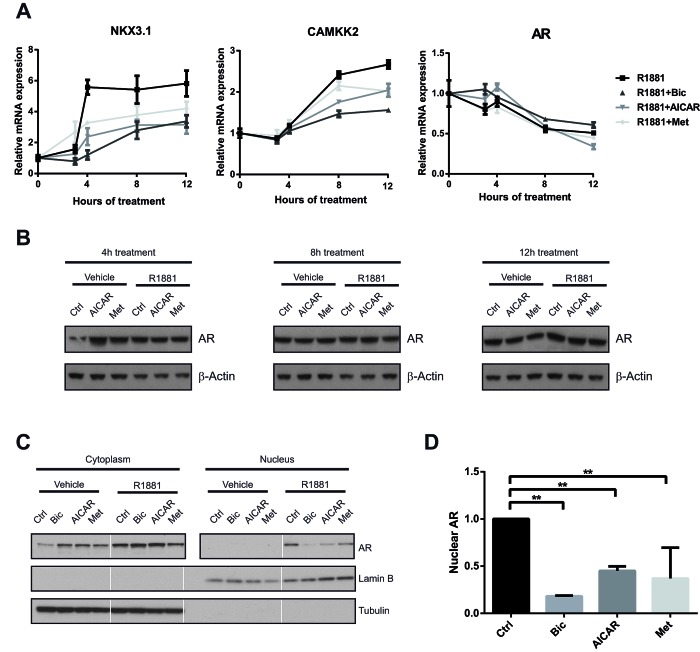
Figure 5. Activation of AMPK reduces nuclear localisation, but not expression of AR.
A: To assess the dynamics of AR inhibition following AMPK activation, LNCaP cells were grown in androgen-free medium for three days, then stimulated with R1881 and bicalutamide, AICAR or metformin for 3, 4, 8 and 12 h. Expression of NKX3.1, CAMKK2 and AR was quantified by qRT-PCR. n=3. B: To evaluate effects of AICAR and metformin on AR protein level, LNCaP cells were grown in androgen-free medium for three days, then stimulated with R1881 and bicalutamide, AICAR or metformin for 4, 8 and 12 h. AR protein levels were assessed by Western Blot. C: To assess effects of AICAR and metformin on localisation of the AR, C4-2 cells were grown in androgen-free medium and stimulated with drugs as indicated for 12 h. Cytoplasmic and nuclear fractions were separated, and AR protein levels were assessed by Western Blot. Lamin B and tubulin were used as loading controls and to confirm successful fractionation. White lines separate non-contiguous bands run on the same gel. D: Nuclear AR levels after androgen stimulation were quantified using Western Blots from three independent experiments and normalised to the Lamin B signal.

