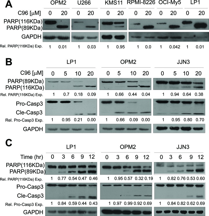
Figure 6. C96 activates apoptotic signaling in MM cells.
(A) OPM2, U266, KMS11, RPMI-8226, OCI-My5, and LP1 cells were treated with C96 (20 μM) for 24 hrs. Cell lysates were then prepared and subjected to immunoblotting assay against apoptosis-associated proteins PARP, and apoptotic executive enzyme pro-caspase-3 (Pro-Casp3). GAPDH was used as a loading control. (B) LP1, OPM2, and JJN3 were treated with C96 at the indicated concentrations for 12 hrs, followed by immunoblotting assay for pro-caspase-3 (Pro-Casp3), cleaved caspase-3 (Cle-Casp3), and PARP. GAPDH was used as a loading control. (C) LP1, OPM2, and JJN3 were treated with C96 at 10 μM for different time points, followed by immunoblotting assay for caspase-3 and PARP. GAPDH was used as a loading control.

