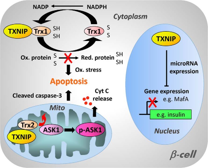
Figure 1.
Schematic diagram of the cellular functions of TXNIP. In the cytoplasm, TXNIP binds to and inhibits thioredoxin 1 (Trx1) and thereby interferes with the ability of Trx1 to reduce oxidized proteins, resulting in oxidative stress and increased susceptibility to apoptosis. In addition, TXNIP can also enter the mitochondria where it interacts with mitochondrial thioredoxin 2 (Trx2), releasing ASK1 from its inhibition by Trx2 and allowing for phosphorylation and activation of ASK1. This in turn leads to cytochrome c (Cyt C) release from the mitochondria, cleavage of caspase-3, and apoptosis. TXNIP has also been found to be localized in the nucleus and to modulate the expression of various microRNAs (eg, miR-204). These microRNAs down-regulate the expression of target genes including important β-cell transcription factors such as MafA, which results in reduced insulin transcription and impaired β-cell function.

