Figure 3.
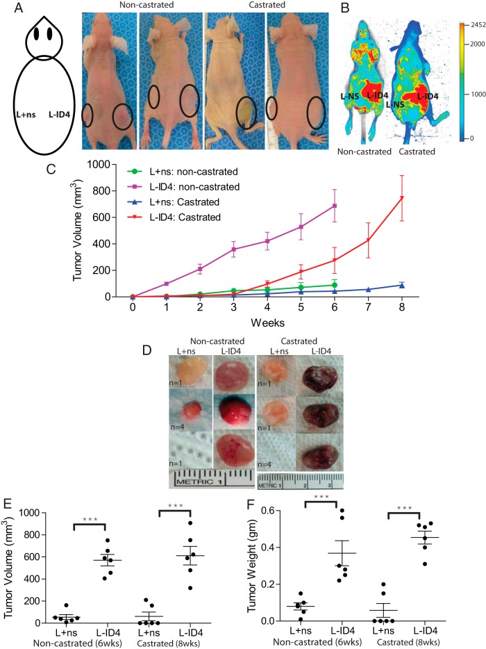
Inactivation of ID4 in LNCaP cells promotes tumor growth in vivo. A, L+ns and L-ID4 cells in Matrigel were injected into the left and right flanks of nude mice (male nu/nu), respectively, as shown in the schematic diagram. Tumor growth is shown by solid circles. B, At the end of the experiments, mice were injected with IRDye 800CW endothelial growth factor receptor targeting agent and evaluated for tumor-specific retention of the fluorophore by near-infrared fluorescence imaging (red). C, Volumes of the tumors were measured weekly (expressed as cubic millimeters, means ± SEM, n = 6/group). The noncastrated mice were killed at 6 weeks, whereas castrated mice were killed at 8 weeks. D, Representative xenograft images with numbers of mice with similar tumor (n) are shown. E and F, Respective volumes and weights (means ± SEM, n = 6) of the tumors after excision from the mice (***, P < .001, between L+ns and L-ID4 tumors in noncastrated and castrated mice, respectively).

