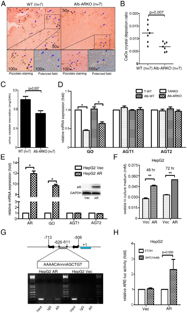
Figure 3.
Mice lacking AR in hepatocytes had suppressed oxalate synthesis via down-regulated GO activity. A, Crystal staining showing the deposition areas of CaOx crystals in kidney tissues of the Alb-ARKO and their WT littermate control mice. CaOx crystal formation was detected by Pizzolato staining and polarized light optical microphotography. Arrowheads indicate CaOx crystals. B, Quantitation of CaOx crystals in each kidney section. Higher numbers of CaOx crystals were found in the WT mice than in the Alb-ARKO mice. C, Detection of 24-hour oxalate excretion in urine samples of the WT and Alb-ARKO mice. D, mRNA expressions of the oxalate synthesis–related enzymes (GO, AGT1, and AGT2) in liver tissues of the TARKO and Alb-ARKO mice and their WT littermate (T-WT and Alb-WT, respectively) control mice. E, mRNA expressions of the enzymes GO, AGT1, and AGT2 upon manipulation of the AR level in human liver carcinoma HepG2 cells (Western blot in the right corner shows AR knockin efficiency). *, P < .05). F, Oxalate level measurement in HepG2 cell culture media. AR promoted oxalate synthesis in HepG2 cells. **, P < .01. G, ChIP assay using the GO promoter. AR increased binding on the −626 to −611 region of the GO promoter. H, Luciferase assay. A luciferase (luc) construct containing the AR binding site of the GO promoter region was designed. AR promoted luciferase activity–containing AR binding sites in the GO promoter region. ETOH, ethanol; DHT, dihydrotestosterone; Vec, vector.

