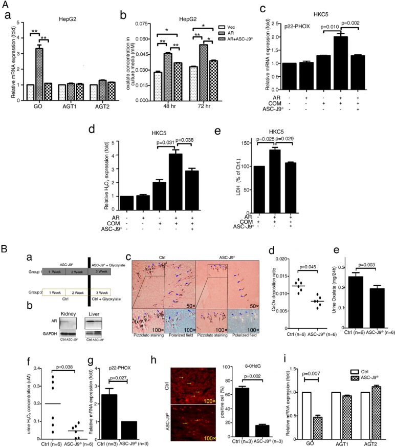
Figure 6.
ASC-J9 lowered CaOx crystal formation in mouse kidney via targeting AR. A, In vitro studies: ASC-J9 effect on expressions of oxalate synthesis-related enzymes (GO, AGT1, and AGT2) (a), oxalate excretion in culture media (b), NADPH oxidase subunit p22-PHOX expression (c), H2O2 production (d), and LDH release (e). The HepG2 and HKC5 cells, AR-incorporated and vector control cells, were used in all assays. *, P < .05; **, P < .01. B, In vivo studies, a, A diagram describing the injection schedule for ASC-J9. b, Western blot showing AR degradation in liver and kidney tissues of mice. Mice were injected ip with ASC-J9 (75 mg/kg) every day for 3 weeks. Control mice were injected with vehicle, N,N-dimethylacetamide. c, Crystal staining showing the deposition area of CaOx crystals in kidney tissues of the ASC-J9-treated and vehicle control mice. CaOx crystal formation was detected by Pizzolato staining and polarized light optical microphotography. Arrowheads indicate CaOx crystals. d, Quantitation of CaOx crystals was reduced by treatment with ASC-J9. e, 24-hour oxalate excretion in urine of the ASC-J9—treated and the vehicle control mice. The ASC-J9–treated mice showed decreased urine oxalate excretion compared with that of the vehicle-treated control mice. f, Detection of urine H2O2 levels in the ASC-J9–treated mice and the vehicle control mice. The ASC-J9–treated mice showed lower H2O2 levels. g, NADPH oxidase subunit p22-PHOX mRNA expression levels in the kidney tissues of the ASC-J9–treated mice and the vehicle control mice. The ASC-J9–treated mice showed decreased NADPH oxidase subunit p22-PHOX mRNA expression. h, IF staining of 8-OHdG in ASC-J9–treated mice showing that loss of AR decreased the number of 8-OHdG positively stained cells in the mouse kidney (quantitation on the right). i, qPCR assay showing GO, AGT1, and AGT2 mRNA expression in ASC-J9–treated mouse liver tissues. CTRL, control.

