
Figure 7.
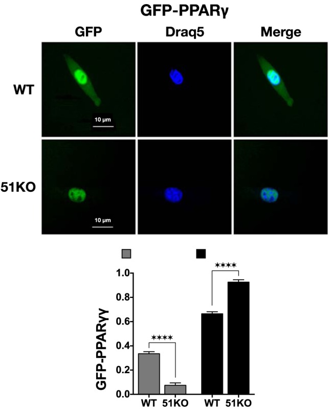
FKPB51 regulates steady-state intracellular localization of PPARγ to the cytoplasm. WT and 51KO MEFs were transfected with PPARγ-GFP in media containing charcoal-stripped serum to remove free fatty acids. DRAQ5 was used to label nuclear DNA. GFP and DRAQ5 were visualized 24 hours after transfection using a Leica DMIRE2 confocal microscope. Images shown are representative of 3 independent experiments where a minimum of 50 cells per condition were inspected. The fluorescence microscopy images were used with ImageJ to measure cellular localization of PPARγ-GFP. All results represent means ± SEM (n = 9). ****, P > .0001.