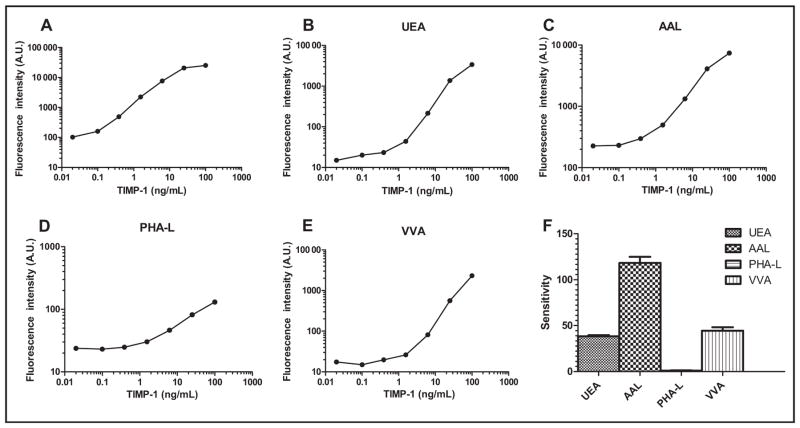
Fig. 2. (A) Establishment of the integrated system for protein quantification and glycan detection in TIMP-1: magnetic bead–based immunoassay for TIMP-1 protein; (B–E), dose–response curves of UEA, AAL, PHA-L, and VVA immunosorbent assays for TIMP-1; F, glycan profile of the recombinant TIMP-1.
The glycan profile was established using the sensitivity of the dose–response curves, calculated as changes of fluorescence intensity per TIMP-1 concentration. A.U., arbitrary units.

