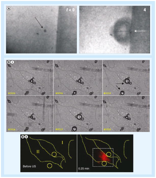
Figure 2. Microbubbles undergo cavitation in contact of cells.
(A) Formation of microjet. Before ultrasound exposure (left), the arrow points to a microbubble held in place by an optical trap. During ultrasound exposure (4 μs later; right), the microbubble collapses, and the arrow points to a microjet directed toward the cell attached to the surface on the right. (B) Series of high-speed images show (i) microbubbles (arrows) experience stable cavitation (oscillations) during ultrasound exposure that (ii) led to cellular uptake of the cell impermeable reagent propidium iodide.
US: Ultrasound.
(A) Reproduced with permission from [8]; (B) Reproduced with permission from [41].

