
Fig. 1. Mechanical development of heart and brain tissue parallels expression of abundant cell and matrix proteins.
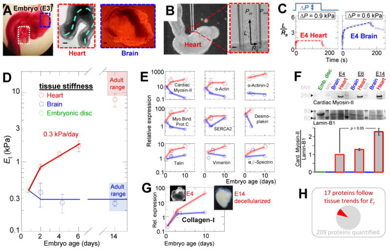
(A) E3 chick embryo with heart tube (white box) and midbrain (blue box) in situ and after isolation. The heart continues to beat ex vivo, with contraction and flow propagating along the dashed line. Scale = 100 μm. (B) Micropipette aspiration of an E3 heart tube in phase contrast microscopy. Scale = 10 μm. (C) Representative aspiration and relaxation curves for E4 heart and brain demonstrate the respective elastic and inelastic responses. (D) Et for heart and brain tissue throughout embryonic development, starting with day-1 embryonic disk, then E2, E4, E6, and E14 heart and brain (n ≥3 measurements each). By the time beating starts, the heart is already 3-fold stiffer than early embryonic tissue and then stiffens at a rate of 0.3 kPa/day. Due to the thick epicardium of E6 hearts and older relative to the inner diameter of our micropipettes, measurements likely underestimate stiffness of the myocardium at those stages due to significant contribution of epicardium. Brain tissue does not stiffen during development and remains viscoelastic with a mean Et = 0.3 kPa. (E) Quantitative mass spectrometry (MS) of cellular proteins extracted from intact embryonic disc (Hamburger-Hamilton stage 3-4), E2, E3, E4, and E10 heart and brain tissue reveals a small set of detected proteins with expression patterns similar to heart or brain mechanics: namely, a general increase in heart and relatively small increase in brain. Expression is relative to average in brain E2-E3 (n≥3 MS measurements). (F) Immunoblot confirms that MS measurements of Cardiac Myosin-II expression increase in heart development. Samples are pooled from 3-4 embryos at each reported stage and were normalized to Lamin-B1 (n ≥3). (G) MS indicates that collagen-I expression increases during heart development, but not greatly during brain development. Inset images: 1% SDS-decellularized E4 and E14 hearts. The insoluble matrices retain the shape of the embryonic hearts, but while E14 matrix (with 80% of MS ion current being collagen-1) appears solid, the E4 matrix appears more reticulated and porous, consistent with relatively less mass. (H) Of the proteins identified by Mass-Spec (Table S1), a small subset had expression levels across tissues and development that paralleled mechanics. Error bars in all figures represent SEM.