Figure 6.
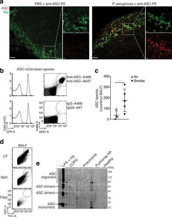
Extracellular ASC specks are formed in vivo and accumulate during human chronic inflammatory disease. (a) Confocal imaging of subcapsular sinus macrophages of popliteal draining LNs from P. aeruginosa infected mice injected 4 h later via the same route with 4 μg of PE-labeled anti-ASC Abs. Scale bars: 53 μm left panels 37 μm right panels, 9 μm inserts. (b) Flow cytometry of ASC-mCerulean specks pre-incubated with two monoclonal anti-ASC Abs (top histograms) or IgG1 isotype controls (bottom histograms) directly labeled with A488 and A647 fluorophores. Amount of specks per μl of sample was determined by subtracting A488+A647+ events in anti-ASC stained sample from those in IgG1 control stained samples. (c) Adjusted quantification of A488+A647+ ASC specks in cell-free BALF from WT BALB/c mice exposed to cigarette smoke or normal air for 8 weeks. (d) Flow cytometry of BALF samples from patients with COPD (5 ml/patient) before and after removal of dead cells by centrifugation (400 x g, 5 min) and size filtration (5 μm). (e) Immunoblotting for ASC (right) of cell-free filtered BALF samples from patients with COPD (n = 4), Pneumonia (n = 4), Pulmunary hypertension (n =2) or healthy donors (n =2) after chemical crosslinking with 1 mM of DSS. Cell-free sups from untreated (–) or LPS-primed, nigericin-activated THP-1s were used as controls. Data show representative images (a) flow cytometry (b, d), and immunoblotting (e) analysis from one out of two independent experiments; (c) cumulative from one experiment; each symbol represents an individual mouse; horizontal and vertical lines indicate mean + SD, *P = 0.0317, Mann-Whitney test.

