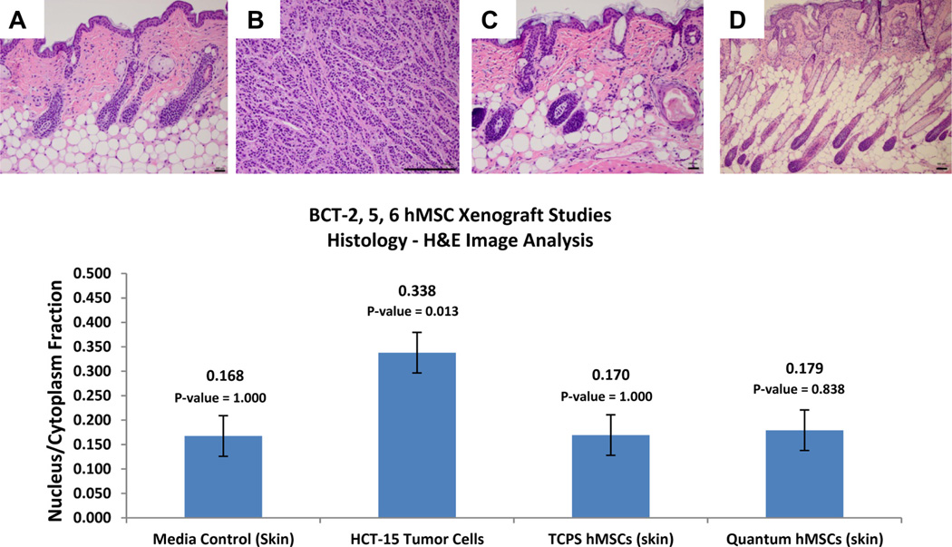
Figure 9.
Xenograft hematoxylin and eosin histology. (A–D) Representative sections of host-animal tumor and injection site dermal tissue for BCT-2, BCT-5 and BCT-6 experiments. Groups A (negative control; α-MEM-based cell culture media), C (TCPS-derived BM-hMSCs) and D (Quantum-derived BM-hMSCs) are negative for malignant neoplasm in the three experiments. Only group B (containing HCT-15 tumor cells) arms were positive for malignant neoplasm. Image analysis of hematoxylin and eosin histology sections from all three xenograft studies is also shown. Nucleus/cytoplasm ratios were computed from LABCAT digital processing software. These data indicate that there is no significant difference between the nucleus/cytoplasm ratios of host animal tissue implanted with BM-hMSCs expanded in either TCPS or the Quantum. Student t test P values for HCT-15 tumor cells, TCPS BM-hMSCs and Quantum BM-hMSCs all were comparisons with the media control group results. (Scale bars = 100 µm.)

