Figure 2.
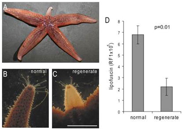
Asterias rubens with an arm regenerating from the base (A–C). The early regenerate (C) is less pigmented than an intact arm of the same animal (B). Fluorometrical analysis of extracted lipofuscin content shows lower levels of lipofuscin in regenerating arms than in the tips of corresponding intact arms of the same animals (D). Lipofuscin is expressed as mean+/− s.e.m. Relative fluorescence intensities (RFI). R, regenerate; N, normal/intact arm. Light micrographs and data were obtained from animals with approximately 1 cm regenerates. Data obtained from animals with 1.5–2 cm regenerates. Scale bar: 1 cm (B, C). From (Hernroth et al., 2010).

