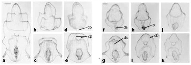
Figure 3.

Bipinnaria larvae of P. ochraceus at various stages of regeneration after surgical bisection. The anterior larva is shown in the top row, and the posterior larva in the bottom row. a, Bipinnaria before surgical treatment. b, c, Day 1. Anterior (b) and posterior (c) larvae after surgical division. The gut of the posterior larva is intact enough for feeding. d, Day 3. e, Day 4. Elongation along the larval axis has taken place. f, g, Day 7. The anterior larva (f) has regenerated a functional digestive system. h, Day 9. i, Day 10. Complete regeneration of ciliary bands has occurred in both anterior and posterior larvae. j, k, Day 12. Both larvae have regenerated and are similar to control larvae. cb, ciliary band; cp, coelomic pouch; ds, digestive system; p, phytoplankton. a–e, Scale bar (in a), 200 μm. f–k, Scale bar (in f), 200 μm. From (Vickery and McClintock, 1998)
